Clear Sky Science · en
Stage-Dependent mediation of white matter hyperintensities between plasma biomarkers and cognitive function in Alzheimer’s disease
Why Brain Spots Matter for Everyday Thinking
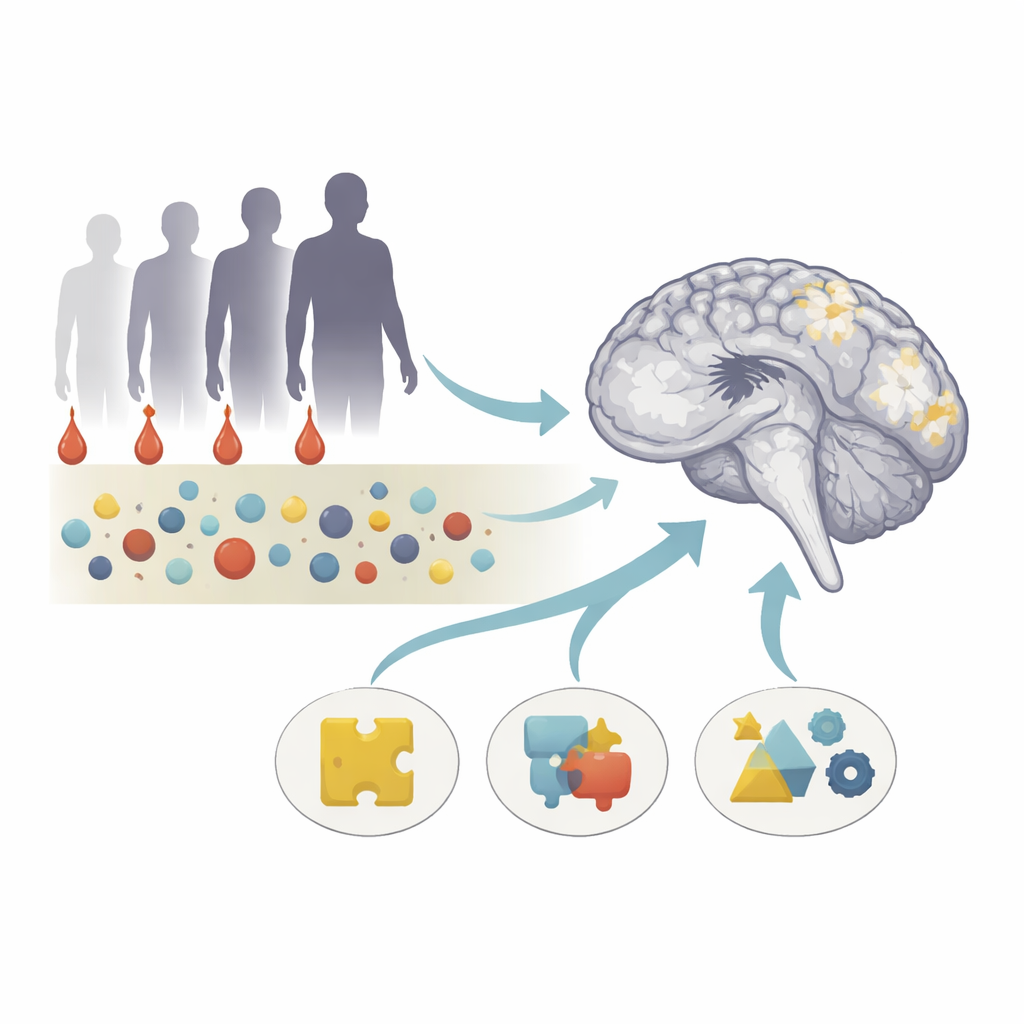
Alzheimer’s disease is usually linked to sticky protein clumps and shrinking brain tissue, but doctors also see bright “spots” in brain scans of many older adults. These spots, called white matter hyperintensities, mark areas where the brain’s wiring may be damaged. This study asked a pressing question: how do simple blood tests and these bright spots together explain who is likely to develop memory and thinking problems, and how this changes from healthy aging to full Alzheimer’s disease?
Following People Across the Memory Spectrum
The researchers studied 311 volunteers ranging from healthy older adults to people with subjective memory complaints, mild cognitive impairment, and diagnosed Alzheimer’s disease. Everyone completed a detailed set of thinking and memory tests, had a high-resolution brain MRI scan, and gave a small blood sample. Instead of treating all bright spots in the brain as the same, the team divided them into four regions based on where they sit relative to the brain’s fluid-filled spaces and outer surface. This allowed them to ask whether specific locations of damage are more closely tied to blood changes and different types of thinking problems.
Blood Clues from Amyloid and Nerve Damage
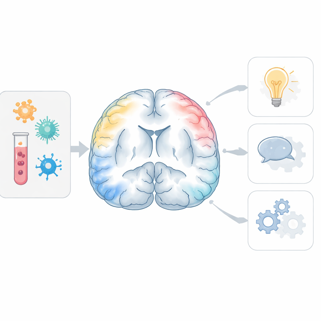
In the blood, the team measured a ratio of two forms of amyloid protein (Aβ42/Aβ40), which signals how much Alzheimer-type change is happening in the brain. They also tracked glial fibrillary acidic protein, reflecting inflammation in support cells, and neurofilament light chain, a marker of injury to long nerve fibers. As expected, people with Alzheimer’s had lower amyloid ratios and higher levels of the two damage markers than cognitively healthy participants. Lower amyloid ratio was linked to poorer overall thinking scores and to weaker memory, language, and higher-level planning skills, showing that a simple blood draw can capture subtle cognitive decline.
Bright Spots in Critical Brain Wiring
Brain scans revealed that not all white matter spots are equal. Compared with healthy and mildly affected individuals, people with Alzheimer’s had much larger clusters of bright spots close to the brain’s fluid spaces and just under the cortex, while deeper areas were less clearly involved. Larger volumes in these key regions were tied to worse scores on global tests and on specific abilities such as memory, language, and executive function. Importantly, lower amyloid ratio and higher inflammation and nerve-injury markers went hand in hand with more of these region-specific spots, even after accounting for age, education, and common vascular risks like high blood pressure and diabetes. This suggests that Alzheimer-related biology directly contributes to damage in the brain’s wiring, beyond classic blood vessel disease.
A Shifting Pathway from Protein to Thinking Problems
The most striking finding was how these relationships changed with disease stage. In people who were still cognitively normal, the bright spots closest to the brain’s fluid spaces appeared to carry part of the impact of abnormal amyloid on thinking, especially on language and overall mental status. In other words, amyloid changes in blood were linked to subtle thinking problems partly because they were associated with these early wiring changes. In people who were already cognitively impaired, the picture grew more complex: the same regions, along with nearby periventricular areas, also reflected inflammation and nerve fiber injury, and these combined changes explained a larger share of the link between blood markers and cognitive decline.
What This Means for Patients and Prevention
To a lay observer, this work suggests that a particular pattern of bright spots around the brain’s fluid spaces may be an early bridge between abnormal Alzheimer blood markers and later problems with thinking, memory, and language. Early in the disease, amyloid changes alone seem to disturb these sensitive regions, while later on, added inflammation and nerve damage join in, turning a single-path problem into a multi-faceted cascade. By combining blood tests with careful mapping of where white matter damage occurs, clinicians may one day better predict who is on a fast track to cognitive decline and tailor treatments to the stage of disease—focusing on amyloid and vascular health early, and adding anti-inflammatory and nerve-protective strategies as symptoms advance.
Citation: Chen, H.J., Guo, Y., Huang, W. et al. Stage-Dependent mediation of white matter hyperintensities between plasma biomarkers and cognitive function in Alzheimer’s disease. Transl Psychiatry 16, 140 (2026). https://doi.org/10.1038/s41398-026-03927-5
Keywords: Alzheimer’s disease, white matter hyperintensities, brain MRI, blood biomarkers, cognitive decline