Most people bounce back after a frightening experience, but for some, reminders of the event trigger intense fear and vivid memories for months or years. This condition, known as post-traumatic stress disorder (PTSD), is closely tied to how the brain stores and updates memories of danger. The hippocampus—a seahorse-shaped brain region crucial for forming and recalling contexts and events—often shows shrinkage and abnormal activity in people with PTSD. This study uses mice to explore a simple question with big implications: how do stress hormones, released right after trauma, change hippocampal cells in ways that can lock in unhealthy fear and disrupt normal memory?
Building a Better Trauma Model in Mice
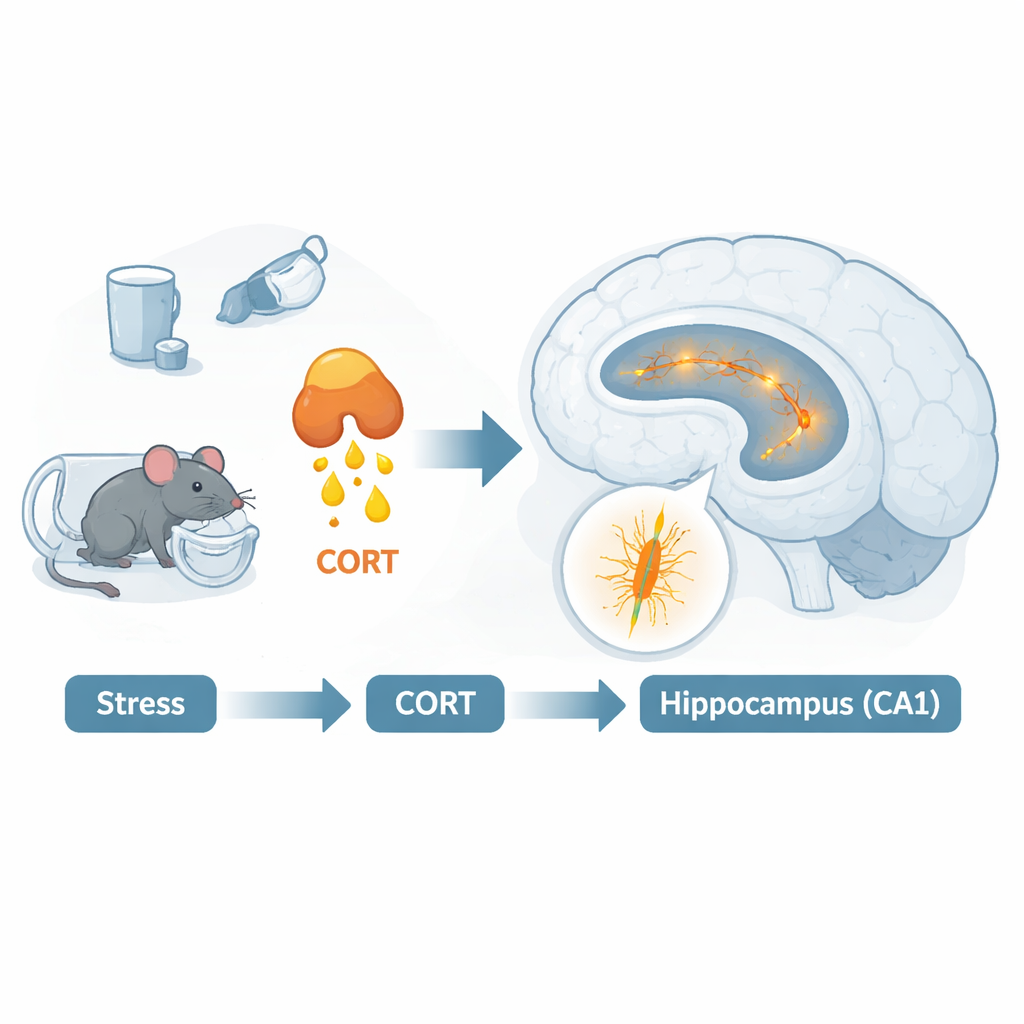
Researchers often rely on a protocol called single prolonged stress (SPS) to mimic aspects of PTSD in rodents. It combines several intense stressors—such as restraint, forced swimming, and brief anesthesia—and has produced reliable PTSD-like effects in rats. In mice, however, the results are inconsistent: some strains show strong changes in fear and memory, others do not, suggesting a hidden vulnerability that only shows up under certain conditions. The authors suspected that stress hormones themselves, especially corticosterone (the rodent equivalent of cortisol in humans), might be one such missing factor. They designed a model in which young adult male mice received SPS followed immediately by an injection of corticosterone, aiming to more closely mirror the hormone surge that follows a traumatic event in people.
After SPS and a 10-day recovery period, the mice went through a series of behavioral tests. In an open field arena, stressed animals with or without added hormone showed normal movement and no obvious increase in anxiety-like behavior. But in a Y-shaped maze that probes short-term spatial working memory, the SPS-plus-corticosterone mice performed worse: they were less likely to alternate between arms in a flexible pattern and more likely to repeat visits to the same arm. Next, the animals were trained in a contextual fear task, where the environment—not a tone—predicts a mild foot shock. All groups learned the association, but only the SPS-plus-corticosterone mice later showed “contextual amnesia”: they froze less when returned to the shock-paired setting, as if the environment no longer strongly signaled danger. At the same time, these animals struggled to extinguish fear over repeated safe re-exposures, a hallmark of PTSD-like behavior.
How a Single Channel Quieted Memory Neurons Figure 2.
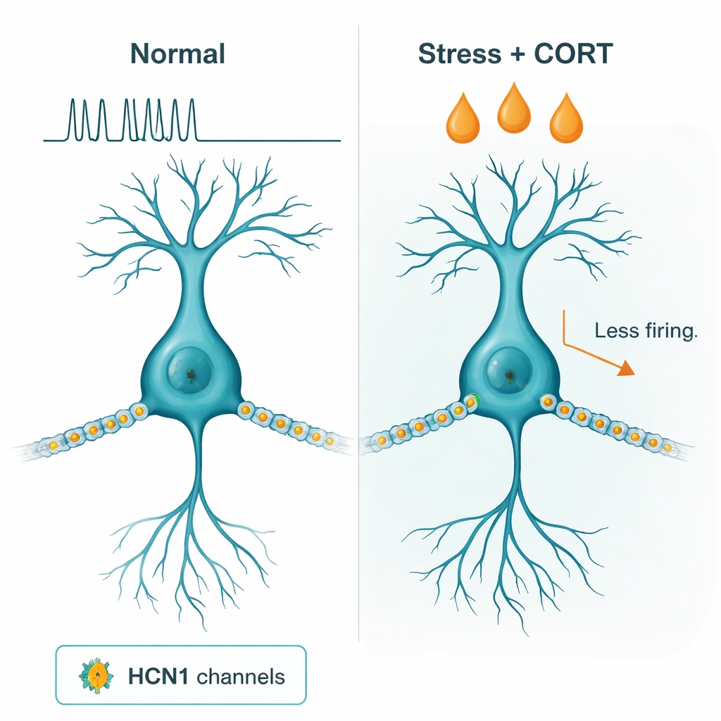
To understand what was happening inside the hippocampus, the team made thin brain slices and recorded electrical activity from individual neurons in the dorsal CA1 region, an area central to spatial and contextual memory. In mice that experienced SPS plus corticosterone, these cells were harder to excite: they had lower input resistance and fired fewer action potentials in response to current. The researchers traced this change to an increase in a particular electrical current, called Ih, which flows through proteins known as HCN1 channels. In SPS-plus-corticosterone mice, Ih was larger and activated more easily, meaning these channels opened at less negative voltages and acted like powerful leaks that shunted incoming signals. When the scientists applied a drug that blocks HCN channels, the neurons’ electrical properties returned to normal, and their ability to fire in response to input was restored.
Proving Cause and Effect with Genetic Tweaks
Correlation alone was not enough; the authors wanted to know whether HCN1 was truly driving the behavioral changes. They used viruses to either boost or delete HCN1 specifically in dorsal CA1 pyramidal neurons. Overexpressing HCN1 in stressed mice, even without extra hormone, was sufficient to reproduce the key features seen in SPS-plus-corticosterone animals: poorer spatial working memory, weaker recall of the fear-associated context, and difficulty extinguishing fear. Electrophysiological recordings confirmed that these neurons resembled those from the hormone-treated group, with reduced excitability and elevated Ih. Conversely, when HCN1 was selectively removed in CA1 neurons of SPS-plus-corticosterone mice, their memory performance improved and neuronal excitability normalized. In other words, the channel was both sufficient to generate the deficits and necessary for them to appear.
Why This Matters for Trauma and Treatment
For non-specialists, the key message is that this study links a specific molecular “valve” in memory cells—HCN1 channels—to the way traumatic stress and stress hormones combine to distort memories. In this mouse model, SPS alone did not reliably produce PTSD-like problems, but adding a burst of corticosterone uncovered a persistent weakness in the hippocampus: its neurons became too quiet to properly encode and update contextual fear. By showing that turning HCN1 up or down can worsen or rescue these deficits, the work identifies a concrete target for future drugs aimed at easing memory-related symptoms of PTSD. While much remains to be tested in other ages, sexes, and brain regions, the findings suggest that carefully dialing hippocampal excitability—rather than simply dampening fear responses—could be a promising route to more precise treatments after trauma.
Citation: Kim, C.S., Kim, J. & Michael, S. Effects of post-stress corticosterone on hippocampal excitability and behavior involving hyperpolarization-activated cation channel 1 function.
Transl Psychiatry16, 74 (2026). https://doi.org/10.1038/s41398-026-03871-4