Clear Sky Science · en
Aberrant dynamic functional architecture in major depressive disorder: Vertex-Wise large-sample fMRI analyses reveal network-specific alterations and symptom associations
Why this brain study on depression matters
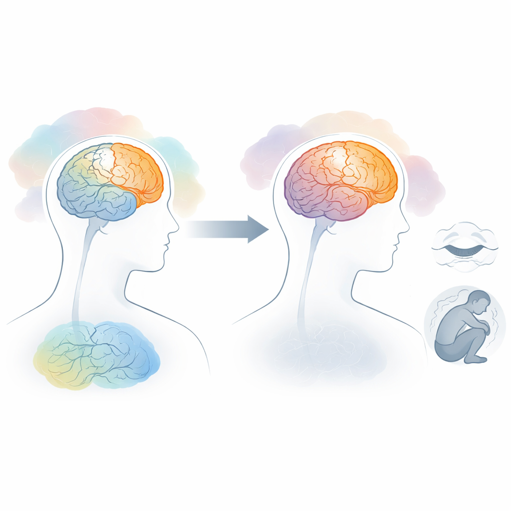
Major depression touches hundreds of millions of people worldwide, yet we still do not fully understand what is going wrong in the brain. Most brain scans used in research take a kind of long-exposure “snapshot” of activity and miss how the brain’s communication patterns change from moment to moment. This study uses a large collection of brain scans and a newer way of looking at brain activity over time to show that depression is linked to a disturbed balance between brain areas that handle inner thoughts and those that process the outside world. The work also ties specific brain changes to symptoms like insomnia, guilt, and reduced insight, hinting at future, more tailored treatments.
A moving picture of brain activity
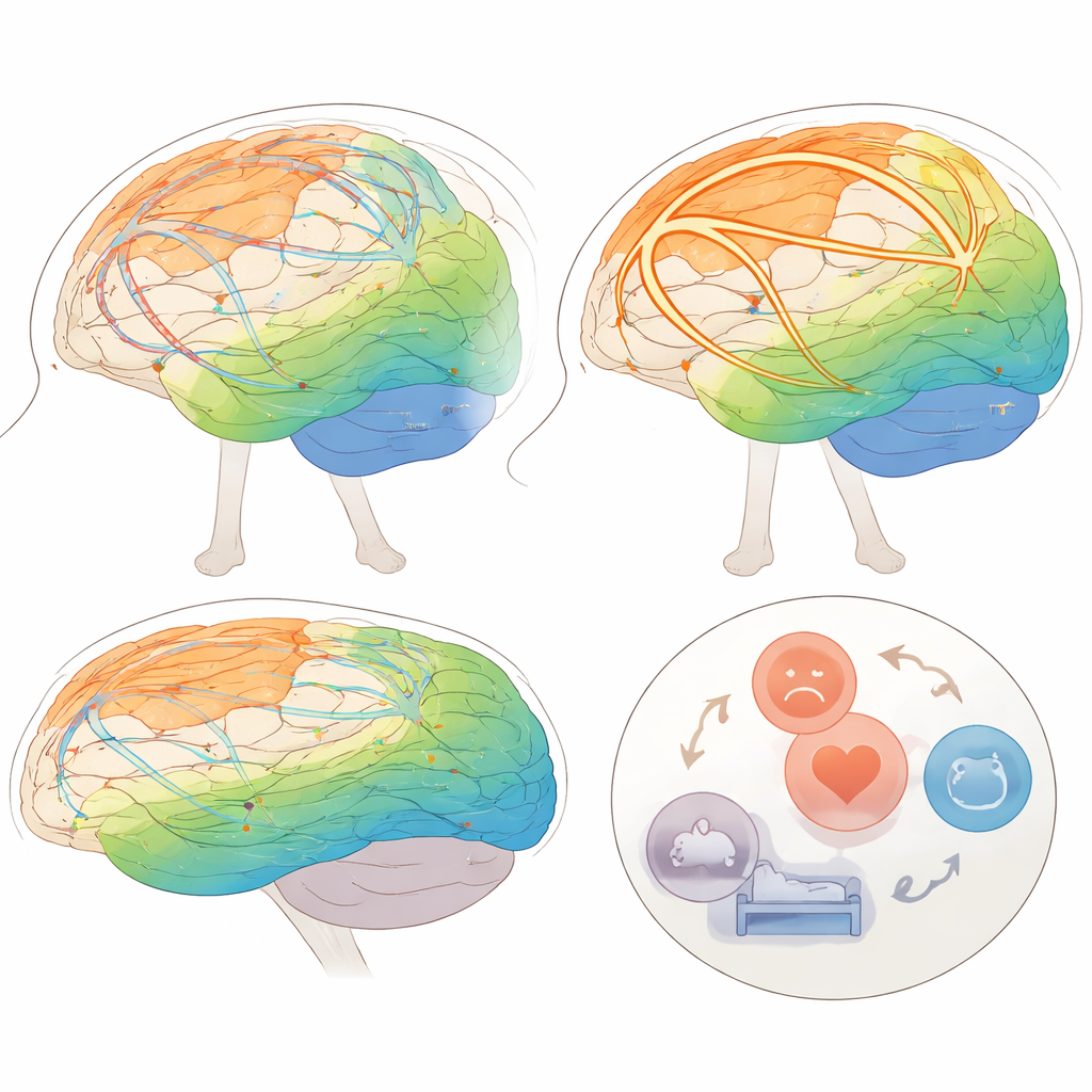
Traditional brain imaging studies in depression focus on static connectivity, averaging signals over several minutes and treating brain networks as if they were fixed. Yet brain activity is constantly shifting even when we rest quietly in a scanner. In this study, researchers analyzed resting-state fMRI data from nearly 3,000 people, including over 1,500 with major depressive disorder and more than 1,300 healthy volunteers, collected by the Depression Imaging Research Consortium (DIRECT). Instead of averaging everything together, they slid a time window along each person’s scan and examined how strongly each point on the brain’s surface communicated with all other points from one window to the next. This allowed them to compute a “temporal stability” score—how consistently each region stays in a similar communication pattern over time.
Where the brain grows too rigid and too fragile
Across all participants, both healthy and depressed, the brain showed a common overall pattern. Higher-order regions involved in complex thinking and self-reflection—such as frontal and parietal areas and parts of the so-called default mode network—tended to have more stable communication patterns. Primary sensory and motor regions, which must rapidly react to sights, sounds, and bodily signals, were more changeable. In people with depression, however, this balance was shifted. The study found increased stability in many higher-order “association” regions, including frontal control areas and limbic regions that help regulate emotion, while stability dropped in primary sensory and motor regions and in parts of the visual system. In simple terms, networks that support inwardly focused thought became more locked-in, whereas systems that connect us to the external world became less reliably organized.
Linking brain dynamics to everyday symptoms
To connect these brain changes to lived experience, the researchers related temporal stability in key regions to detailed clinical ratings of depressive symptoms. They highlighted a set of areas—the superior frontal regions, parts of the postcentral (body-sensing) cortex, and the superior insula (a hub for bodily feelings and internal awareness)—that showed altered stability and were associated with specific complaints. For example, abnormal stability in the superior frontal cortex was tied to feelings of guilt and sleep-onset problems, while changes in the postcentral gyrus and insula related to different types of insomnia and insight into illness. Follow-up analyses of how these regions flexibly connect with the rest of the brain over time revealed a common disturbed network linking frontal, sensory-motor, cingulate, and insular areas, suggesting that difficulties in switching between internal focus and external engagement may underlie clusters of symptoms.
What this could mean for treatment
The study’s large sample, fine-grained surface-based methods, and focus on moment-to-moment dynamics give weight to its conclusions. Rather than one single “depression network,” the results point to an imbalance: overly rigid inner-focused networks and unstable sensory-motor systems that together may fuel rumination, disturbed sleep, and distorted self-perception. These findings dovetail with brain targets already used in treatments like transcranial magnetic stimulation and deep brain stimulation. By sharpening our picture of which dynamic patterns relate to which symptom clusters, such work may help move care toward more personalized interventions that aim not just to change how strongly regions are connected, but how flexibly those connections reconfigure over time.
Citation: Li, XY., Lu, B., Chen, X. et al. Aberrant dynamic functional architecture in major depressive disorder: Vertex-Wise large-sample fMRI analyses reveal network-specific alterations and symptom associations. Transl Psychiatry 16, 127 (2026). https://doi.org/10.1038/s41398-026-03812-1
Keywords: major depressive disorder, brain connectivity, resting-state fMRI, dynamic networks, insomnia