Clear Sky Science · en
Oxytocin neurons in the anterior and posterior paraventricular nucleus have distinct behavioral functions and electrophysiological profiles
Why one “bonding” chemical can push us together or apart
Oxytocin is often called the “love hormone” because of its reputation for strengthening social bonds, from parent–infant care to romantic attachment. But in both humans and animals, oxytocin can also heighten anxiety and fuel social withdrawal after stress. This study asks a deceptively simple question: are there different groups of oxytocin-producing brain cells that separately drive friendly approach versus wary avoidance? By mapping and manipulating these cell groups in mice, the authors show that where oxytocin is made in the brain can flip its effects from comforting to cautionary.
Two neighborhoods in the brain’s social hub
Oxytocin is produced in a small but influential brain region called the paraventricular nucleus (PVN), which sits in the hypothalamus and sends signals to many other social and emotional centers. The researchers focused on two parts of this region: an anterior (front) zone and a posterior (back) zone. They first charted where oxytocin-producing neurons lie along this front–back axis in both California mice (a monogamous, highly social species used to study stress) and standard laboratory mice. They then compared these PVN neurons to a third group of oxytocin cells located in the bed nucleus of the stria terminalis (BNST), a region previously linked to stress-induced social vigilance and avoidance. 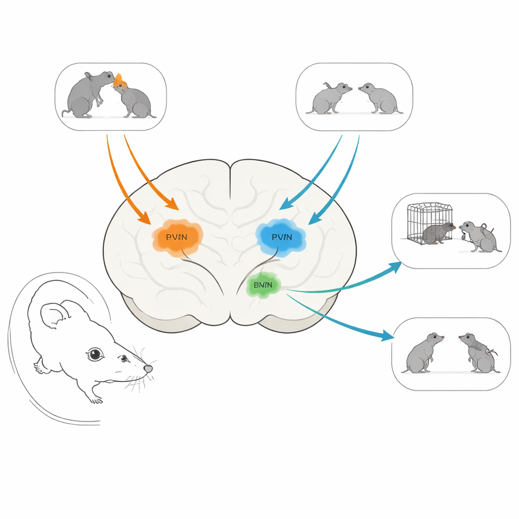
Stress, social behavior, and turning down oxytocin
In California mice, social defeat stress—repeated exposure to an aggressive peer—causes long-lasting social withdrawal and watchfulness in females. Earlier work showed that this kind of stress especially boosts activity in oxytocin neurons in the front PVN of females. To test what these neurons actually do, the team used a molecular “off switch” called a morpholino to reduce oxytocin production in either the anterior or posterior PVN. When they knocked down oxytocin in the front PVN of females that had experienced defeat, the usual stress effects were weakened: these mice spent more time approaching a new mouse and showed less scanning, vigilant behavior. Importantly, turning down oxytocin in this region did not change general movement or interest in an empty cage, suggesting a specific role in how animals respond to social stress rather than in overall activity or curiosity.
A different role for oxytocin further back
The back part of the PVN told a very different story. When the researchers reduced oxytocin in the posterior PVN of unstressed male and female California mice, those animals became less willing to approach a new mouse, even though their vigilance, movement, and exploration of an empty cage were unchanged. In other words, oxytocin from the posterior PVN seemed to support normal, friendly social engagement in both sexes under baseline conditions. Together, these experiments reveal that two nearby oxytocin pools in the same brain structure promote opposite social strategies: the anterior group helps drive avoidance and caution after stress, while the posterior group encourages approach.
Zooming in on how these neurons behave
To understand why these cell groups behave differently, the team recorded the electrical properties of individual oxytocin neurons in anterior PVN, posterior PVN, and BNST of genetically engineered mice whose oxytocin cells glow under the microscope. Neurons in the anterior PVN and BNST were more “excitable”: they received more frequent synaptic inputs and fired more spikes in response to injected current than posterior PVN neurons. Posterior PVN oxytocin cells, by contrast, had fewer but larger synaptic events and a more negative resting state, making them less likely to fire quickly. Despite these differences, all three oxytocin groups received a mix of excitatory and inhibitory inputs. The pattern suggests that anterior PVN and BNST oxytocin neurons form a high-gain, readily activated network suited to rapidly shaping defensive social responses, whereas posterior PVN neurons operate as a calmer, slower system that may reinforce positive social contact. 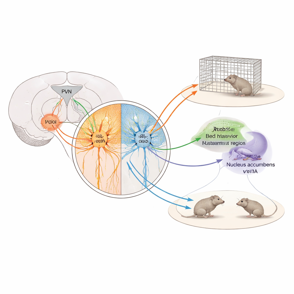
What this means for understanding oxytocin’s double life
This work shows that oxytocin is not a single “good” or “bad” social chemical, but a family of circuits with distinct jobs. Cells in the front PVN and in the BNST help animals become wary and keep their distance after threatening encounters, potentially improving survival in hostile environments. Cells in the back PVN, in contrast, appear to support ordinary social approach, likely by engaging reward-related brain regions. For people hoping to use oxytocin-like drugs to treat anxiety, autism, or trauma, these findings are a reminder that targeting oxytocin broadly could strengthen both comforting bonds and fearful avoidance. Future therapies may need to home in on specific oxytocin pathways—rather than simply boosting oxytocin everywhere—to nudge social behavior in the desired direction.
Citation: Chrisman, A.N., Sugimoto, C., Butler-Struben, H. et al. Oxytocin neurons in the anterior and posterior paraventricular nucleus have distinct behavioral functions and electrophysiological profiles. Neuropsychopharmacol. 51, 946–955 (2026). https://doi.org/10.1038/s41386-026-02352-y
Keywords: oxytocin, social behavior, stress, paraventricular nucleus, neural circuits