Clear Sky Science · en
Broadband plasmon modulation and high-intensity nanofocusing for high-resolution nanoscale imaging using Fabry–Pérot probes
Bringing Light to the Nanoscale
Many of today’s most exciting technologies—from next-generation chips to single-molecule biosensors—depend on seeing and probing structures far smaller than the wavelength of light. This paper reports a new kind of ultra-sharp optical fiber probe that squeezes ordinary laser light into a tiny, intensely bright spot only a few dozen nanometers across, opening the door to sharper images and more sensitive measurements at the nanoscale.
A Tiny Needle of Light
Conventional microscopes are limited by diffraction: they cannot resolve details much smaller than about half the wavelength of light. To get around this, researchers use near-field probes that bring light to within a few nanometers of a surface. The device studied here is an optical fiber that tapers down to a metal-coated, needle-like tip. Light travels down the fiber, turns into surface waves on the metal, and concentrates at the apex, creating a nanoscale “flashlight.” These surface waves, called surface plasmon polaritons, can trap light energy in spots much smaller than those possible with normal lenses.
Smarter Design for Stronger Focusing
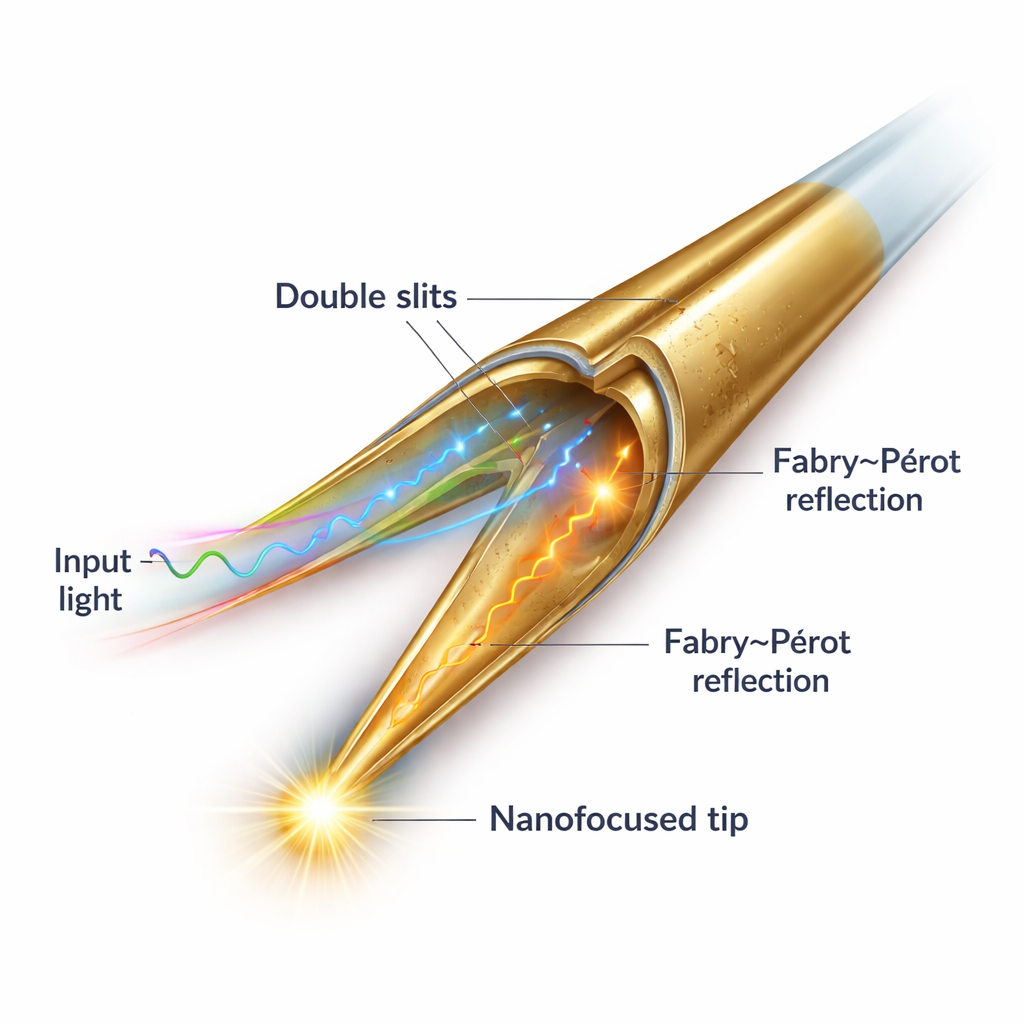
Existing probes face two big hurdles. First, they often require a special, donut-shaped polarization of light that is difficult to produce and very sensitive to alignment. Second, they lose a lot of energy along the way, so the light at the tip is weak and the resulting images are noisy. The authors overcome both problems by building a carefully patterned gold structure on the fiber tip. Two offset half-ring slits carved into the metal act like a tiny polarization control device, converting ordinary linearly polarized light inside the fiber into a symmetric surface wave that can travel efficiently to the very end of the tip without being cut off or spilling into the background.
A Built‑In Light Recycling Cavity
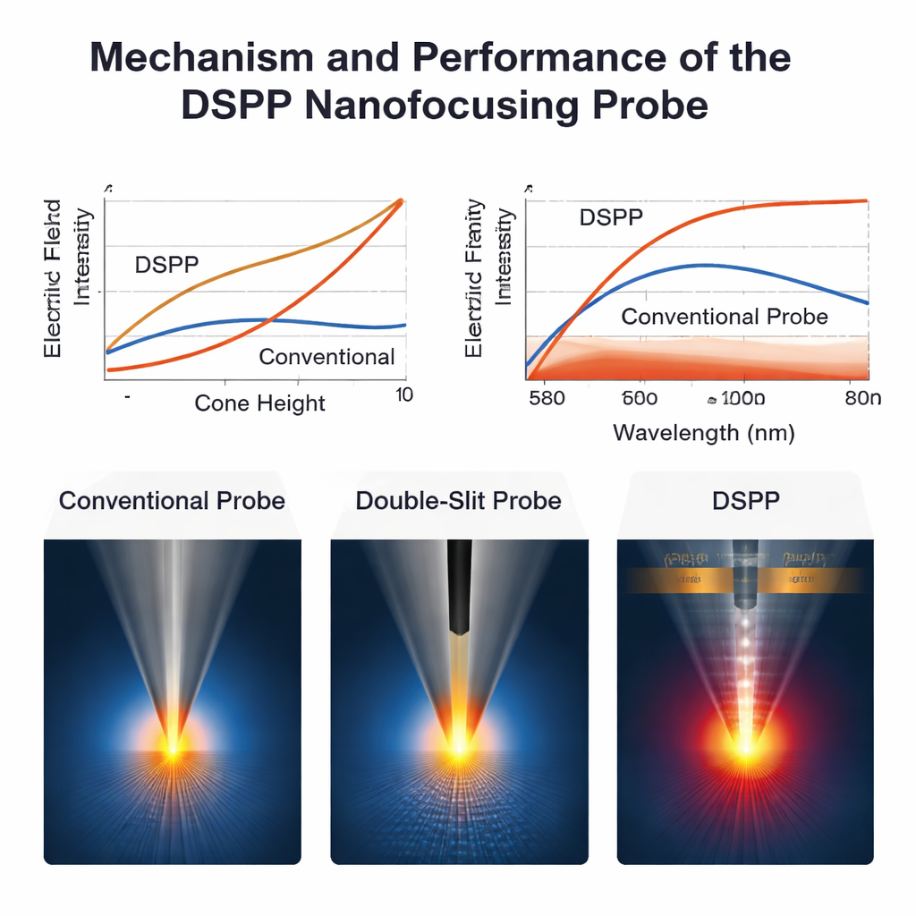
Beneath the sharp apex, the team introduces a flat “platform” region that behaves like a microscopic hall of mirrors for the surface waves. When the waves reach the tip and focus, part of the energy keeps moving past the apex and travels down the opposite side of the cone. There, the flat platform reflects the waves back toward the tip. If the height and angle of the cone are chosen correctly, these returning waves arrive in step with the incoming ones, stacking up like synchronized ripples on a pond. This Fabry–Pérot–like effect greatly boosts the electric field at the tip, leading to a nanofocused spot that simulations and experiments show is about six times stronger than that of an earlier double-slit design under the same illumination.
Sharper, Brighter, and Across Many Colors
To make such a delicate structure practical, the authors develop a “sleeve ring” focused ion beam etching method that lets them sculpt the conical tip and the flat platform with nanometer precision and a tip radius of only about 15 nanometers, far smaller and more repeatable than with traditional chemical etching. They then test how the probe behaves over a wide range of visible wavelengths, from roughly yellow to deep red light. Both simulations and measurements show that the probe maintains a tightly confined hotspot across this broad band, and that its energy recycling design is especially effective at shorter wavelengths, where metal losses are normally most severe.
Imaging Details Smaller Than 30 Nanometers
To demonstrate what this means in practice, the researchers image a gold structure with an extremely narrow slit, just under 30 nanometers wide. Atomic force microscopy and electron microscopy confirm the true shape and size of the slit. Using their new probe in a near-field optical setup, they clearly resolve the slit and the surrounding triangular features, and the measured optical profile gives a width of 28.6 nanometers—showing that the optical resolution rivals that of the mechanical probe and far exceeds what a standard confocal microscope can achieve, which only shows a blurred outline due to the diffraction limit.
Why This Matters
In plain terms, this work delivers a sharper, brighter, and easier-to-use nanoscale flashlight at the end of an optical fiber. By turning simple, linearly polarized light into a strongly concentrated near-field spot and recycling lost energy back to the tip, the new probe design achieves deep subwavelength resolution and strong signals without exotic light sources or fragile alignment. This makes it a powerful candidate for tasks such as examining defects on chips, mapping optical properties of advanced materials, and probing biological structures and molecules one by one, all under ordinary laboratory conditions.
Citation: Dong, H., Hu, W., Ji, P. et al. Broadband plasmon modulation and high-intensity nanofocusing for high-resolution nanoscale imaging using Fabry–Pérot probes. Microsyst Nanoeng 12, 71 (2026). https://doi.org/10.1038/s41378-026-01197-1
Keywords: near-field optical imaging, plasmonic fiber probe, nanofocusing, super-resolution microscopy, nanoscale sensing