Clear Sky Science · en
Super-resolution functional photoacoustic microscopy via label-free cell tracking
Seeing the Brain’s Smallest Blood Highways
The health of our brain depends on countless tiny blood vessels delivering oxygen to hard‑working nerve cells. Until now, scientists could not watch this oxygen traffic in three dimensions at the level of individual red blood cells without adding dyes or labels. This study introduces a new imaging approach that does exactly that, opening the door to clearer insights into how strokes and other brain diseases disturb the brain’s oxygen supply.
A New Way to Listen to Light
The researchers built a microscope based on photoacoustics, a technique where very short bursts of laser light heat light‑absorbing molecules in blood by a tiny amount, causing them to emit ultrasound waves. Instead of using a traditional ultrasound sensor, they created a clear micro‑ring resonator—a tiny optical ring on a transparent chip—that sits on a window in the skull. Laser light passes through this ring into the brain, and the returning ultrasound subtly changes how light circulates in the ring. By reading these changes, the system turns them into detailed pictures of blood vessels and the oxygen carried by red blood cells, all without injecting any contrast dyes. 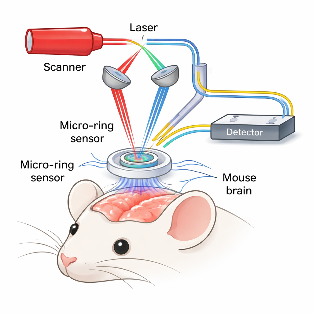
Tracking Single Blood Cells in 3D
Conventional photoacoustic microscopes can clearly separate individual red blood cells from one another when viewed from above, but they blur together along the depth of the tissue. The authors solved this by rapidly repeating thin cross‑section scans through the brain at a thousand frames per second, then digitally tracking the motion of each red blood cell from frame to frame. By following these paths across hundreds of scans, they “connect the dots” into a super‑sharp three‑dimensional map of the microvessel network. At the same time, they use two different colors of laser light to distinguish oxygen‑rich from oxygen‑poor hemoglobin, allowing them to calculate the oxygen level in each tiny vessel segment.
Matching Gold‑Standard Microscopy
To prove that their new method, called super‑resolution functional photoacoustic microscopy (SR‑fPAM), was truly accurate, the team compared it directly with two‑photon microscopy, a powerful but more invasive imaging technique that requires fluorescent dyes. Looking at the same regions of mouse cortex, they found that SR‑fPAM resolved vessels and capillaries with nearly the same fine detail in all three dimensions, down to the scale of single red blood cells. Careful analysis showed that the shapes and positions of vessels in the new images closely matched those from two‑photon imaging, but SR‑fPAM added native information about blood oxygenation and flow direction without extra labeling.
Watching a Tiny Stroke Reshape Blood Flow
The researchers then used SR‑fPAM to watch how the brain’s microvessels respond when a single small artery on the surface is deliberately blocked—a model of a tiny stroke. They could see, in real time, which nearby vessels lost blood flow completely, which ones reversed their flow direction, and how fast the red blood cells moved before and after the blockage. Importantly, they measured how oxygen levels fell in stalled vessels and then recovered as other pathways picked up the slack. The images reveal a complex, three‑dimensional rerouting of blood flow and oxygen delivery, as the brain recruits alternate routes to protect threatened tissue. 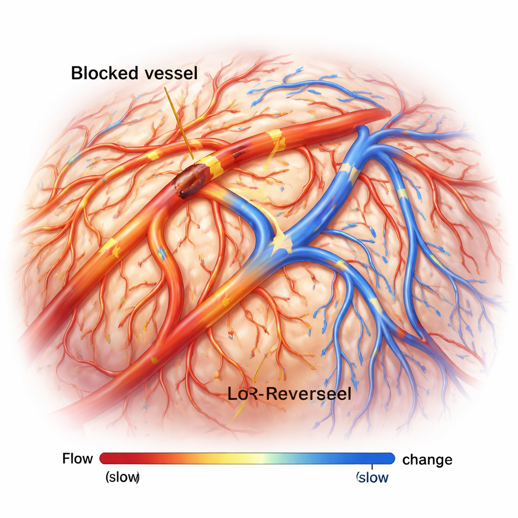
What This Means for Brain Health
By combining label‑free imaging, single‑cell detail, and full three‑dimensional coverage of structure, flow, and oxygenation, SR‑fPAM fills a major gap in how scientists can study the living brain. It offers a way to see not just where blood goes, but how well it carries oxygen through the finest vessels during health, stroke, and other conditions. In the future, pairing this technique with measurements of nerve cell activity could give a far more complete picture of how blood supply and brain function are linked—and how that partnership breaks down in diseases such as stroke, dementia, and hypertension.
Citation: Zhong, F., Wang, Z., Lee, Y. et al. Super-resolution functional photoacoustic microscopy via label-free cell tracking. Light Sci Appl 15, 146 (2026). https://doi.org/10.1038/s41377-026-02235-3
Keywords: photoacoustic microscopy, brain microcirculation, oxygen metabolism, neurovascular coupling, ischemic stroke