Clear Sky Science · en
Large zoom ratio and adaptive aberration correction microscope using 4DPSF-aware Physical Degradation-guided Network
Sharper Views of the Hidden World
Microscopes let us see cells, tissues, and tiny structures that are invisible to the naked eye, but anyone who has used one knows the trade‑offs: you often must swap lenses by hand, refocus, and accept that high zoom can mean dimmer, blurrier pictures. This research presents a new kind of microscope that zooms smoothly over a wide range like a camera lens while an intelligent computer system cleans up the image in real time, promising faster, clearer views for biology, medicine, and materials science.
Why Ordinary Microscopes Fall Short
Conventional lab microscopes change magnification by rotating between fixed objective lenses. That mechanical switching breaks the viewing flow, can cause the image to jump, and limits how quickly scientists can track fast events such as moving cells. New "liquid lenses," whose focus can be changed electrically, offer the hope of smooth zooming. But by themselves they cannot bend light enough for very high magnification, and they introduce complex optical flaws—known as aberrations—that change with zoom level and position in the image, making pictures softer, distorted, or fringed with color.
Smart Optics that Can Really Zoom
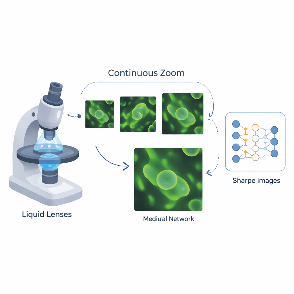
The team designed a continuous-zoom objective for a microscope built around electrowetting liquid lenses, whose curvature changes when a voltage is applied. By arranging liquid and solid lenses in two cooperating groups and adding a movable relay image plane, they created a flexible optical layout that can vary magnification from about 10.6× to over 100× without swapping hardware. Carefully tuned movements keep the sample in focus while both lens groups share the zooming work, extending the usable zoom range beyond what liquid lenses alone could achieve.
Teaching a Network How the Lens Misbehaves
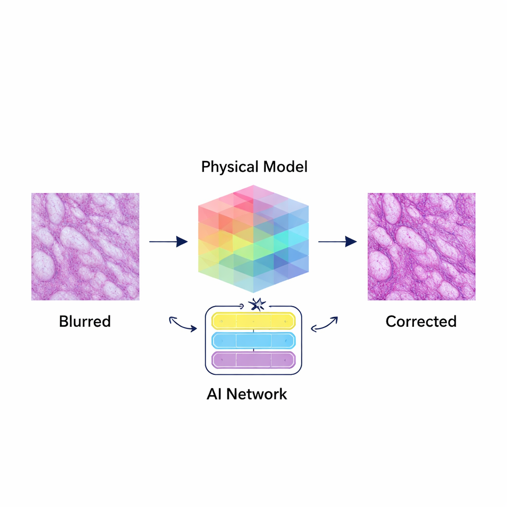
Even with this advanced lens design, image quality can still suffer because the way light spreads and blurs—the point spread function, or PSF—changes across the field of view, with wavelength, and with magnification. Rather than trying to fix these problems after the fact with generic image filters, the researchers built a neural network that is explicitly aware of the optics. They simulated how the microscope blurs light in four dimensions (position, color, and zoom level) and fed this 4D PSF information directly into their 4DPSF-PDNet, a deep learning model that uses a learnable version of a classic deblurring method and an attention-based module to restore details while suppressing noise.
Putting the System to the Test
To prove the idea works, the team first used optical design software to optimize the lens system and generate thousands of paired sharp and degraded images that mimic what the microscope would see at different zoom levels. They then trained their network both on these simulations and on real microscopic pictures taken from a variety of biological samples and compared its performance with other state-of-the-art image restoration methods. Across multiple magnifications, especially under strong optical flaws, their approach produced clearer, more accurate images, improving a standard quality measure (peak signal-to-noise ratio) by about 2.5 to 3 decibels over leading competitors. Tests on resolution patterns and tissue slices, such as sections of small intestine, showed that the microscope can smoothly zoom while keeping structures centered and sharply resolved.
What This Means for Future Microscopy
For a non-specialist, the main takeaway is that this work marries a smart, electrically tunable lens system with an equally smart correction algorithm that knows the physics of the optics. Together they deliver smooth, camera-like zooming in a microscope without sacrificing detail, and they automatically clean up blur and color errors that would normally limit what scientists can see. Such an adaptive system could help pathologists scan tissue slides more quickly, allow cell biologists to follow tiny processes across scales, and aid materials researchers in examining defects, all while reducing the need for manual lens changes and refocusing.
Citation: Yu, DX., Jiang, Z., Zheng, Y. et al. Large zoom ratio and adaptive aberration correction microscope using 4DPSF-aware Physical Degradation-guided Network. Light Sci Appl 15, 140 (2026). https://doi.org/10.1038/s41377-025-02155-8
Keywords: adaptive microscopy, liquid lens zoom, image aberration correction, physics-guided deep learning, biological imaging