Clear Sky Science · en
Speckle-based X-ray microtomography via preconditioned Wirtinger flow
Seeing Inside Objects with Gentle X-rays
X-ray scans are powerful for peering inside objects, from fossils to food and biological tissue. But many everyday materials barely absorb X-rays, so standard scans can miss fine details or require multiple exposures and more radiation. This paper introduces a new way to capture rich, three-dimensional internal structure from a single X-ray snapshot, using a smart mathematical technique called preconditioned Wirtinger flow (PWF) and a simple sheet of sandpaper as a diffuser.
Turning Random Graininess into Useful Information
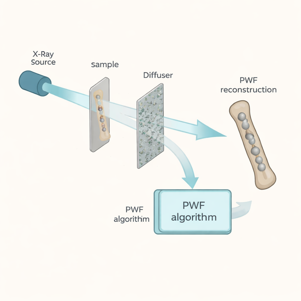
Instead of trying to form a sharp shadow image, the researchers deliberately create a grainy, mottled pattern called “speckle.” In their setup, a beam of hard X-rays passes through the sample, then through a thin random diffuser (such as stacked fine sandpaper) before reaching the detector. The sample subtly shifts and distorts this speckle pattern. Hidden in those tiny shifts is information about how the X-rays slowed down and dimmed as they traveled through the sample, which is closely related to the material’s internal structure and composition.
Recovering Phase Without Extra Assumptions
For materials that do not absorb X-rays strongly—like soft tissue, wood, or many polymers—the most revealing quantity is not how much the beam is dimmed, but how much its wavefront is delayed, known as the “phase.” Existing speckle-based techniques typically estimate only the local bending, or gradient, of this phase and often rely on repeated measurements with the diffuser moved to multiple positions, plus simplifying assumptions about the sample. PWF, in contrast, works from a single speckle measurement and a separate reference image taken without the sample. It uses a physics-based model of how X-rays propagate, interact with the diffuser, and blur due to the partial coherence of the source—important for both synchrotron and compact laboratory X-ray systems.
Smart Algorithms for Finer Details
The heart of the method is an iterative mathematical engine that starts with a guess of the sample’s complex field—how much the wave is attenuated and phase-shifted at each point—and repeatedly refines this guess so that the simulated speckle pattern matches the measured one. A key innovation is a “preconditioner” that steers the updates toward the kinds of changes the speckle image is most sensitive to, namely variations in the phase gradient. A second ingredient, a regularizer based on an oversampling criterion, ensures that there are enough measured speckle grains relative to the unknowns to pin down a unique and stable solution, while naturally limiting how much fine detail can be trusted in the final reconstruction.
Sharper 3D Maps from Fewer X-ray Shots
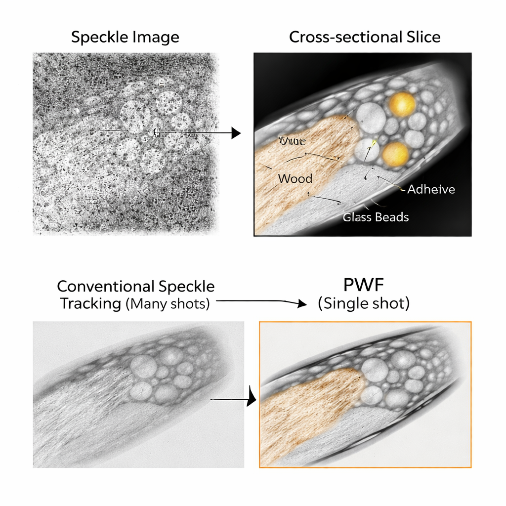
To test their approach, the team imaged a toothpick studded with tiny glass beads, a challenging sample with very large phase shifts and fine internal structure. They compared PWF to one of the best existing “implicit tracking” methods, which needed 12 different speckle images with the diffuser moved each time. Even though PWF used only a single speckle image per viewing angle, it produced three-dimensional maps of the sample’s refractive index that were closer to known values for the glass beads and showed clearer boundaries and fewer artifacts. The method could even recover some information that would normally be treated as diffuse “dark-field” scattering, effectively pushing the resolution down to around 1.5 micrometers in their setup—fine enough to resolve small cellular and microstructural features.
Ready for Real-World Samples
Beyond carefully prepared test objects, the researchers also scanned everyday specimens: a cumin seed, dried shrimp, dried anchovy, and cork. Using the same hardware and reconstruction settings, PWF revealed intricate internal structures and subtle variations in density that are hard to see with conventional absorption-based imaging alone. Because it needs only one speckle pattern per projection angle and already accounts for realistic source blur, the technique promises shorter scan times, lower radiation dose, and simpler hardware. For non-destructive testing, materials science, and potentially even future medical imaging, this work shows that a touch of randomness in the beam, paired with powerful reconstruction algorithms, can turn noisy-looking X-ray images into precise three-dimensional maps of what lies within.
Citation: Lee, K., Hugonnet, H., Lim, JH. et al. Speckle-based X-ray microtomography via preconditioned Wirtinger flow. Light Sci Appl 15, 121 (2026). https://doi.org/10.1038/s41377-025-02118-z
Keywords: X-ray phase contrast, speckle imaging, microtomography, computational imaging, non-destructive testing