Clear Sky Science · en
CSF1R marks a subset of foetal haematopoietic multipotent progenitor cells with acute myeloid leukaemia propagation properties
Why this research matters for babies with leukemia
Leukemia in very young babies is rare but often devastating, and many infants do not respond well to today’s treatments. This study asks a deceptively simple question with big consequences: which early blood cells in the embryo first go wrong to spark one particularly aggressive form of infant leukemia, and can we find a clear handle on them for targeted therapies?
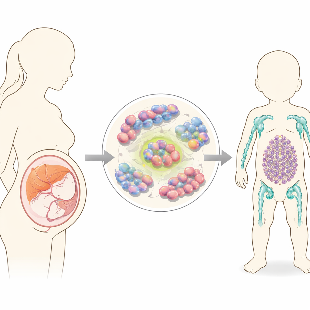
The trouble starts before birth
Doctors have long suspected that many infant leukemias begin in the womb. Clues come from blood spots taken at birth and from twins who share the same cancer-causing mutations. During early development, blood formation moves through several organs, including the yolk sac and a region near the aorta, before settling in the fetal liver and, later, the bone marrow. At each site, different types of immature blood cells appear and disappear. One common genetic accident in infant leukemia is a break and rejoining of a gene called KMT2A with partners such as MLLT3. This rearrangement can drive either a lymphoid leukemia (similar to childhood acute lymphoblastic leukemia) or a myeloid leukemia (acute myeloid leukemia), but it has not been clear which exact fetal cells are first transformed, or what steers them toward one disease type over the other.
A special fetal blood precursor with a myeloid tilt
The researchers focused on a fetal liver population called lymphoid-primed multipotent progenitors (LMPPs). These are early blood cells that can still produce both lymphoid and myeloid lineages. Within this group, they homed in on a subset that carries a surface protein called CSF1R, a sensor for growth signals usually linked to the myeloid branch. Using a mouse model in which the KMT2A::MLLT3 fusion can be switched on during fetal life, they compared CSF1R-positive and CSF1R-negative LMPPs. In lab dishes, both could make lymphoid colonies, but CSF1R-positive cells formed significantly more colonies under myeloid conditions and more often produced “mixed” colonies with features of both myeloid and lymphoid cells, hinting at especially high flexibility and transformative potential.
From flexible precursor to leukemia engine
To test what happens in a living organism, the team transplanted these altered fetal cells into immunodeficient mice. Animals receiving KMT2A::MLLT3-expressing CSF1R-positive LMPPs developed aggressive acute myeloid leukemia: their blood, bone marrow, spleen, liver and even central nervous system filled with immature myeloid blasts, and the disease could be passed on to new mice using bone marrow from the first set of sick animals. In contrast, mice given CSF1R-negative LMPPs initially showed mostly immature B cells in the blood, more reminiscent of lymphoid disease, and took longer to become ill. Genomic analyses revealed that CSF1R-positive LMPPs carried a “stem-like” gene program associated with self-renewal and with known leukemia stem cells in pediatric patients, whereas CSF1R-negative cells showed signatures closer to acute lymphoblastic leukemia.
Survival tricks: self-cleaning and growth signals
The team then asked what allows these CSF1R-marked fetal cells to thrive and drive disease. They found that genes involved in autophagy—cells’ internal recycling and self-cleaning system—were more active in the CSF1R-positive cells. Blocking autophagy with a drug sharply reduced their ability to form colonies. Interfering with CSF1R signaling itself shifted the balance away from myeloid output and, when combined with autophagy blockade, almost completely wiped out colony growth. Importantly, human leukemia datasets showed that a similar CSF1R-positive LMPP-like population exists only during early human development and that CSF1R and several autophagy-related genes are especially active in KMT2A-rearranged acute myeloid leukemia. In a pediatric leukemia cell line carrying KMT2A::MLLT3, a CSF1R inhibitor triggered substantial cell death, supporting the idea that these cells remain dependent on this pathway.
From fetal origin to future therapies
Putting the pieces together, the study suggests that a transient, CSF1R-marked fetal blood progenitor is a likely starting point and engine for KMT2A::MLLT3-driven infant acute myeloid leukemia. These cells combine stem-like endurance with a built-in myeloid bias and rely on both CSF1R signals and autophagy to expand and maintain leukemia. Because CSF1R is a surface molecule already being explored for targeted therapies, including engineered CAR-T cells in adults, this work points toward a concrete, biologically grounded target that could be adapted for vulnerable infants whose disease begins long before they are born.
Citation: Camiolo, G., González Silvera, D., Leah, T. et al. CSF1R marks a subset of foetal haematopoietic multipotent progenitor cells with acute myeloid leukaemia propagation properties. Leukemia 40, 540–552 (2026). https://doi.org/10.1038/s41375-025-02856-4
Keywords: infant leukemia, acute myeloid leukemia, fetal blood development, CSF1R, leukemia stem cells