Clear Sky Science · en
Non-invasive quantitative investigation of varnish stratigraphy in historical artifacts using line-field confocal OCT
Seeing Beneath the Shine
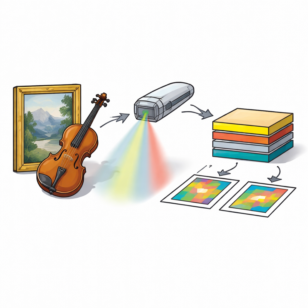
From old master paintings to legendary violins, a clear, glossy varnish is often the final touch that shapes what we see—and how long these treasures survive. Yet these transparent coatings age, are replaced, and sometimes badly overworked, leaving conservators with a delicate puzzle: which shiny layer is original and must be protected, and which can safely be removed? This study introduces a new, non-invasive way to look through those transparent skins in three dimensions, helping experts restore artworks and instruments with far greater confidence.
Why Layers of Shine Matter
Varnish on artworks and wooden instruments is more than cosmetic makeup. It deepens colors, adds gloss, and protects fragile paint and wood underneath. Over centuries, however, these coatings can yellow, crack, or become cloudy. Conservators often strip away degraded, later varnishes and apply new ones, while trying at all costs to preserve any original layer that carries the maker’s intent and the object’s history. The problem is that different varnish coats, glazes, and touch-ups can stack into a complex sandwich only a few hundredths of a millimeter thick. Looking at the surface alone rarely reveals how many layers are present, how thick they are, or which parts were added during past restorations.
A New Way to Look Inside Without Touching
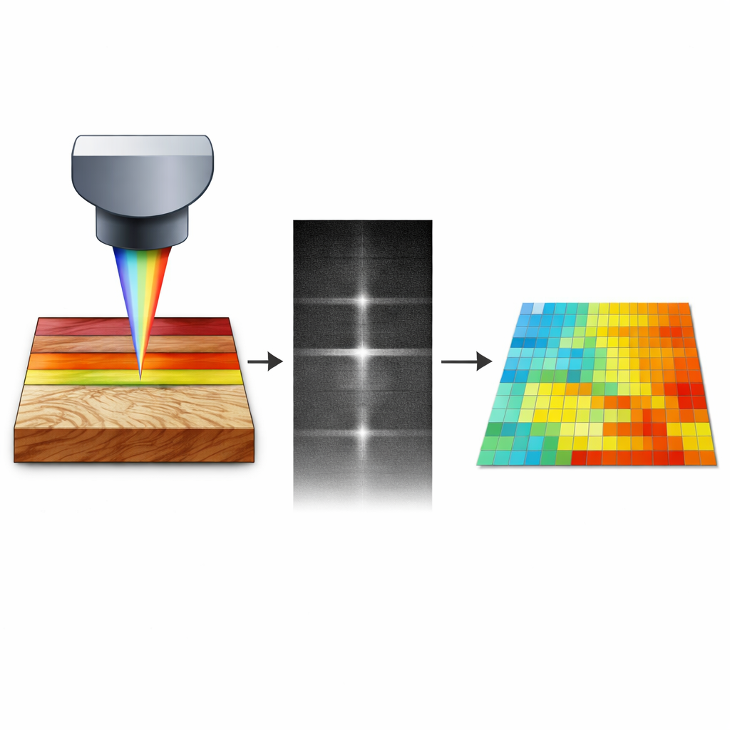
To tackle this challenge, the researchers adapted a medical imaging method called line-field confocal optical coherence tomography (LC-OCT) to the world of cultural heritage. In simple terms, the technique sends a thin line of light into a surface and measures light that bounces back from just below it, building up a high-resolution, slice-like picture through the depth of the material. Unlike traditional microscopes that require taking a physical chip from a painting or a violin, LC-OCT works without contact and can be brought directly to museums or workshops. The team designed a compact, transportable probe, mounted on flexible stands, that can scan paintings on an easel or violins on a bench while capturing 3D views with micrometer-scale detail—fine enough to see individual varnish layers and even tiny filler particles.
Turning Complex Images into Clear Guidance
Raw LC-OCT images look like delicate grayscale cross sections, but they can be hard to interpret by eye alone. The team therefore created open-source software that automatically detects the boundaries between layers and calculates their thickness across an entire scanned volume. The program filters edges, homes in on the key interfaces, then converts the results into colorful thickness maps and statistical charts. This turns an intricate optical signal into clear, quantitative information: where layers begin and end, how uniform they are, and how much varnish remains after cleaning tests. For conservators, this means they can objectively judge whether a solvent or cleaning gel is thinning a coating evenly, leaving residues, or risking the underlying original finish.
Stories from a Damaged Painting and a Famous Violin
The method was put to the test on two very different 17th-century artifacts. On the Spanish painting Notre-Dame del Pilar, LC-OCT revealed where a deep, older varnish still survived beneath a more recent one, and where overpaint had been added to disguise losses. By combining these depth-resolved images with ultraviolet and infrared photography, the conservator could map areas with a single modern varnish, areas with two stacked varnishes, and zones where semi-transparent retouching sat between them. On a 1678 violin by Nicolo Amati, the technique distinguished the original, thick, clear varnish from a later, strongly colored coating applied in the 2000s. Guided by these 3D views, restorers tested mild cleaning mixtures in chosen spots, checking after each step that the intrusive modern varnish was mostly gone while a thin, protective residue and the prized original layer remained intact.
What This Means for Preserving the Past
The study shows that LC-OCT can act as an "X-ray for varnish"—not to identify exact chemistry, but to reveal structure, thickness, and hidden additions with remarkable precision and without removing a single chip. When paired with the trained eye and historical knowledge of conservators and curators, it offers a powerful decision-making tool: where to clean, how far to go, and when to stop. Over time, such non-invasive, quantitative imaging could become a standard part of conservation practice, helping safeguard both the beauty and the authenticity of paintings, instruments, and other varnished treasures for generations to come.
Citation: Galante, G., Vilbert, M., Desvois, L. et al. Non-invasive quantitative investigation of varnish stratigraphy in historical artifacts using line-field confocal OCT. npj Herit. Sci. 14, 193 (2026). https://doi.org/10.1038/s40494-026-02460-4
Keywords: varnish, art conservation, optical coherence tomography, historical paintings, violins